Radius and ulna anterior view – The radius and ulna, located in the forearm, play a crucial role in movement and stability. This anterior view exploration delves into their anatomical features, providing a comprehensive understanding of these essential bones.
The anterior surface of the radius and ulna exhibits distinct shapes, landmarks, and articulations. An organized HTML table showcases these features, facilitating easy comprehension.
Radius and Ulna: General Overview
Anatomical Location and Orientation
The radius and ulna are two long bones that form the forearm. The radius is located on the thumb side of the forearm, while the ulna is on the little finger side. The proximal ends of the radius and ulna articulate with the humerus at the elbow joint, and the distal ends articulate with the carpal bones at the wrist joint.
The radius and ulna are connected by the interosseous membrane, which helps to stabilize the forearm.
Role in Forearm Movement and Stability
The radius and ulna work together to allow a wide range of motion in the forearm, including pronation (turning the palm down) and supination (turning the palm up). The radius also rotates around the ulna during forearm movement, which allows for a greater range of motion.
The ulna provides stability to the forearm and helps to prevent the radius from dislocating.
Anterior View of the Radius and Ulna

The anterior surface of the radius and ulna presents several notable features, including distinct shapes, prominent landmarks, and important articulations. Understanding these aspects is crucial for comprehending the overall anatomy and function of the forearm.
Shape and Landmarks
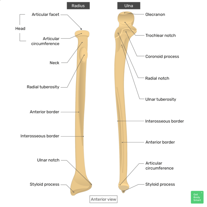
The radius and ulna exhibit unique shapes and landmarks that contribute to their overall form. The radius is characterized by its slender, cylindrical shape, while the ulna is broader and triangular in appearance.
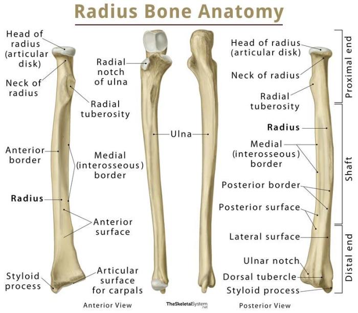
Along the anterior surface of the radius, the following landmarks are readily identifiable:
- Radial tuberosity: A rounded prominence located at the proximal end, which serves as the insertion point for the biceps brachii muscle.
- Bicipital ridge: A vertical ridge extending from the radial tuberosity, providing additional attachment for the biceps brachii.
- Interosseous crest: A sharp ridge running along the lateral margin, serving as the attachment site for the interosseous membrane, which connects the radius and ulna.
The ulna, on the other hand, displays the following prominent landmarks on its anterior surface:
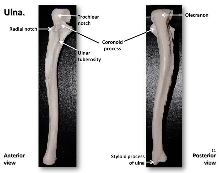
- Trochlear notch: A deep, U-shaped notch located at the proximal end, which articulates with the trochlea of the humerus, forming the elbow joint.
- Coronoid process: A hook-like projection situated above the trochlear notch, which provides additional stability to the elbow joint.
- Radial notch: A shallow notch located on the lateral aspect of the coronoid process, which accommodates the head of the radius.
Articulations
The anterior surface of the radius and ulna facilitates important articulations with neighboring bones.
| Bone | Articulates with | Type of Joint | Function |
|---|---|---|---|
| Radius | Humerus | Pivot joint | Allows rotation of the forearm |
| Ulna | Humerus | Hinge joint | Allows flexion and extension of the forearm |
| Radius and Ulna | Each other | Synovial joint | Permits pronation and supination of the forearm |
Proximal Radioulnar Joint
The proximal radioulnar joint (PRUJ) is a synovial pivot joint formed between the head of the ulna and the radial notch of the radius.
Its articular surfaces are lined with hyaline cartilage, and the joint is surrounded by a joint capsule and various ligaments.
Ligaments
- Annular ligament: Surrounds the head of the ulna and attaches it to the radial notch.
- Quadrate ligament: Connects the head of the ulna to the distal radius and interosseous membrane.
- Radial collateral ligament: Connects the radial notch of the radius to the ulnar shaft.
- Ulnar collateral ligament: Connects the ulnar head to the radius.
Movements, Radius and ulna anterior view
The PRUJ allows for the pronation and supination of the forearm.
- Pronation: Rotation of the forearm so that the palm faces posteriorly.
- Supination: Rotation of the forearm so that the palm faces anteriorly.
Distal Radioulnar Joint

The distal radioulnar joint is a synovial pivot joint that allows for rotation of the distal ulna around the radius. It is located between the distal end of the radius and the ulnar notch of the distal ulna.
The articular surfaces of the distal radioulnar joint are the ulnar notch of the distal radius and the head of the ulna. The joint is stabilized by several ligaments, including the dorsal radioulnar ligament, the palmar radioulnar ligament, and the interosseous radioulnar ligament.
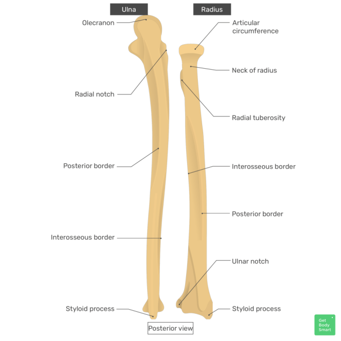
The radius and ulna are two bones in the forearm that are connected at the elbow joint. The radius is located on the thumb side of the forearm, while the ulna is located on the little finger side. The anterior view of the radius and ulna shows the bones from the front.
An archetype is a ________. The radius and ulna are important bones that allow us to rotate our forearm and bend our elbow.
Movements Allowed at the Distal Radioulnar Joint
The distal radioulnar joint allows for rotation of the distal ulna around the radius. This movement is essential for pronation and supination of the forearm.
Clinical Significance: Radius And Ulna Anterior View
The radius and ulna are crucial bones in the forearm, and their anterior view provides valuable information for diagnosing and managing various clinical conditions. Injuries and fractures involving these bones are relatively common, and understanding their clinical significance is essential for healthcare professionals.
Diagnostic imaging techniques such as X-rays, CT scans, and MRIs play a significant role in visualizing the radius and ulna and detecting abnormalities. These techniques help identify fractures, dislocations, and other injuries, guiding appropriate treatment decisions.
Common Injuries and Fractures
- Radius fractures:These are among the most common fractures in the body, often resulting from falls or direct blows to the forearm. Common types include distal radius fractures (wrist fractures) and midshaft radius fractures.
- Ulna fractures:Less common than radius fractures, ulna fractures can occur due to direct trauma or as a result of a radius fracture (known as a Monteggia fracture).
- Distal radioulnar joint injuries:These injuries can involve sprains, strains, or dislocations of the joint, leading to pain, swelling, and limited range of motion.
- Essex-Lopresti injury:A complex injury involving a distal radius fracture and a dislocation of the distal radioulnar joint.
- Radial head fractures:These fractures occur at the proximal end of the radius and can result from a fall or direct impact to the elbow.
Essential FAQs
What is the proximal radioulnar joint?
The proximal radioulnar joint is a pivot joint located at the proximal end of the radius and ulna, allowing for rotation of the forearm.
What movements are allowed at the distal radioulnar joint?
The distal radioulnar joint is a hinge joint that permits flexion and extension of the wrist.